Case Details
INTRODUCTION
In the last 15 years, the evolution of adhesive esthetic materials dramatically modified the restorative approach in the posterior regions, leading to a more and more limited use of the traditional metal restorative materials. Nowadays, if correctly used, the available composite materials and enamel-dentin adhesive systems allow to guarantee very good long-term esthetic and functional results, making esthetic restorations almost invisible in both anterior and posterior regions.
The best advantages offered by resin based materials in comparison to traditional metal alloys regard the esthetic appearance, the preservation of sound tooth structures and the possibility of reinforcing residual tooth structures. Nonetheless, problems regarding polymerization shinkage and dentinal adhesion are still controversial.
Nowadays, the increased reliability of adhesive restorations in the posterior regions is mainly due to hybrid resin composites with mini-particles as well as to the last generation adhesive systems. Such materials are characterized by excellent physical-mechanical properties: in fact, they are densely filled, resistent and radiopaque, their elastic modulus is very similar to that of dentin, they are provided with very good surface characteristics and a wear resistance comparable to those of sound enamel and silver amalgam (10-15 micron/year). Consequently, these materials are indicated for all kinds of restorative cavities.
When a proper quantity of sound enamel is present on all cavity margins, direct adhesive restorations represent the first choice therapy in little and medium I and II class cavities. Different clinical procedures were proposed to compensate for shrinkage stress; the most reliable technique is the segmental stratification using pluri-stratified approaches. The incremental techniques vary according to the cavity type; the most validated are the horizontal, the oblique and the 4-increment technique. Conversely, the traditional three-sites procedure described by Lutz (1986) using transparent matrixes and reflective wedges is no longer used.
In the last decade, several studies showed very satisfactory results using direct adhesive techniques in wide cavities requiring cusp coverage as well.
Nonetheless, in the presence of wide and different cavities without two or more walls, with multiple cusp coverage and with absent or reduced cervical enamel, indirect adhesive restorations (inlays, on lays, overlays) cemented with luting agents should be considered as a viable treatment. Such restorations are stratified on a master model and complete polymerization is achieved in the dental laboratory before intraoral cementation. Consequently, indirect restorations allow to better control polymerization shrinkage, bypass the objective procedural difficulties aimed at restoring a correct morphology of the restoration due to the different steps of direct techniques and are provided with better physical-mechanical properties (better dimensional stability, increased toughness and wear resistance) thanks to post-polymerization procedures.
CASE REPORT
A 27-year old male patient with moderate caries propensity was treated. The patient presented with multiple primary and secondary decays on previous incongruous restorations; such decays were evident at both the clinical and radiographic examination by means of bite-wings.
Some of these decays involved the subgingival part of the teeth.
The treatment plan aimed at establishing proper oral hygiene and periodontal conditions by means of scaling, root planing, polishing and hygienic motivation. Then, a complete esthetic rehabilitation of all intraoral sites was planned using conservative direct and indirect adhesive restorations. Furthermore, a simultaneous periodontal surgica approach was planned to lengthen the clinical crowns of the teeth interested by subgingival lesions. The maxillary third molars were extracted due to their incongruous position, leading to a difficult achievement of a correct home hygienic maintenance; the extraction of such molars did not impair oral functions. Finally, the impacted right mandibular molar was extracted as well.
The orthodontic examination also evidenced a malocclusion but the patient did not accept to treat it.
QUADRANT 1
At level of the right maxillary premolars (teeth 14 and 15), II class adhesive cavity preparations were performed: a rounded occlusal-distal box was designed, the occlusal margins were regularized, the axial walls were slightly rounded while the margin of the cervical box was cut sharp.
In medium II class cavities, the stratification approach of choice is the 4-increment technique, a modification of the “centripetal build-up” proposed by Bichacho in 1994.
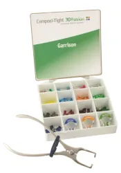
The rubber dam is of paramount importance for the success of the restoration. The sectional matrixes are placed together with the divaricating rings. Such matrixes were specifically developed for adhesive dentistry and allow to achieve effective contact areas.
After applying a three-step etch-and-rinse adhesive system, the marginal crest is built-up with an increment of enamel mass (increment 1); then, the dentin is completely covered with 0.5 mm of a flowable composite resin (increment 2).
The dentin mass is stratified with a unique increment with a maximum thickness of 2 mm (increment 3).
Then, the build-up is completed with the stratification of a unique increment of occlusal enamel (increment 4), modelling the occlusal surface and applying super colors in the occlusal grooves and pits, if necessary. Finally, finishing and polishing of the restorations are performed.
Particularly interesting is the operative sequence proposed for the esthetic and periodontal restoration of the first maxillary molar (tooth 16), in order to recover both the morphological and functional features.
A previous incongruous composite restoration was present with evident microleakage and residual caries that led to a violation of the biological width as well as to inflammation of the soft tissues.
The treatment was planned with a one-step surgical and restorative approach. Once the rubber dam was placed, the restorative material was removed and the carious lesion evidenced. The careful removal of such infiltrated tissues did not allow the rubber dam to properly isolate the operative area, as noticed under the stereomicroscope; moreover, the radiographic control pointed out the invasion of the biologic width.
Consequently, the rubber dam was removed and a lengthening of the clinical crown was performed, in order to restore a correct distance between the sound cervical margin and the bone crest. A resective periodontal surgery procedure was made by means of osteotomy and osteoplasty. In the meanwhile, the third molar was extracted, as previously planed. The flaps were placed in crest with vertical mattress sutures, using 6-0 vicryl sutures.
Thanks to the low rate bleeding due to the vasoconstrictor agent as well as to the rapid and correct surgical procedures, it was possible to place the rubber dam immediately. In order to isolate correctly the post-surgical area, a specific cellulose foam was injected before placing the rubber dam.
Then, the sectional matrix was placed using a Compositigth 3D ring together with a Silver ring. A three-step etch-adn-rinse adhesive system was used. After etching, 2% digluconate chlorexidine was applied for 30 sec in order to inhibit the metalloproteinases and stabilize the adhesive bond over time.
Afterwards, a complex cusp covering restoration was performed with oblique stratification and multiple increments. The first increment was made with a flowable composite resin, in order to fully covering the dentin and to seal the cervical margin in absence of enamel. The marginal crest and the axial mesial-buccal wall were built-up with a double vertical increment of enamel mass; then the oblique stratification of the dentin masses with multiple increments was made; finally, the occlusal surface was modelled with enamel masses and post-polymerization supercolors in the grooves and pits.
The final polymerization was made using a glycerin gel so as to inhibit the superficial oxygen. The restoration was finished and polished as previously described.
The quadrant 1 after the operative procedures and at the 3-month follow-up.
It is worth noticing that the one-step surgical and restorative approach allows to complete the treatment in a single appointment with the following advantages: optimization of the procedural times, elimination of the problems related to the temporary restorations, sealing of the cavity by means of a finished and polished final restoration with correct emergence profiles, rapid and easy healing of the soft tissues.
QUADRANT 2
At level of teeth 24, 25 and 26 II class restorations were made (OD cavities on the premolars and a complex cavity on the first molar). Such restorations were similar to those made on the first quadrant.
It is worth noticing the cavity design on tooth 26, where the non supported cusp was cut by means of an internal bevel, in order to preserve the maximum amount of sound tissue and hold properly the divaricating ring at cervical level. All the three restorations were built up contemporaneously, so as to optimize the operative time. The matrixes were placed forcing adequately the wedges and using the most divaricating rings.
Obviously, the most challenging target was to obtain effective interproximal contact areas together with a correct anatomy on the molar. The segmentation of the stratification was made using the 4- increment technique on the premolars and the oblique stratification technique with multiple increments on the molar with cuspal coverage.
Then, the finishing and polishing procedures were performed, achieving satisfactory morphological and functional results as well as optimal esthetics. Moreover, a good integration of the mesialbuccal cusp of tooth 46 restored with a direct technique was obtained.
At level of tooth 27, an extremely wide occlusal cavity was evident and a microcrack going through the occlusal surface was noticed.
Consequently, two operative choices were available:
a) complete cuspal coverage by means of an adhesively luted overlay;
b) direct restoration preserving the cusps, although interested by the microcrack.
Accordingly to the patient, the latter more conservative option was chosen, even if it was less predictable. The small mesial-occlusal II class was filled with the 3-increment horizontal stratification technique. On the contrary, the wide occlusal cavity affected by the microcrack was restored using the oblique stratifiction technique with multiple increments (flow, 4 increments of dentin, 2 increments of enamel), in order to compensate for the polymerization shrinkage as much as possible.
The final restoration after finishing and polishing and the completed quadrant at the 3-month follow-up, showing proper morphological and functional results as well as good esthetics.
QUADRANT 4
Tooth 47 showed significative loss of tissues on the distal cusps and the decay interested the subgingival area, interfering with the biological width. An incongruous temporary filling was evidenced.
The following treatment plan was chosen: clinical crown lengthening, adhesively luted restoration, extraction of the impacted third molar.
In this case, the author performed a two-appointment combined surgical-restorative procedure, according to the protocol published on the EJED (nr. 1, 2010). A full thickness lingual flap and a double mixed buccal flap were elevated, a limited osteotomy was made and the impacted third molar was extracted.
Then, osteotomy and osteoplasty were performed on tooth 47, lengthening the clinical crown and recovering a proper biological width on the distal surface. The rubber dam was carefully placed, in order to obtain a perfectly isolated area to perform the adhesive procedures.
The adhesive build-up and the cavity preparation were made. The rubber dam was removed and the post-operative impression was immediately taken; although the surgical procedures had been just done, the surgically exposed margins were easily recordable with a correct impression.
After one week (the time necessary to the dental laboratory to build-up the indirect composite restoration), during the second appointment, the sutures were removed and the inlay was intraorally tried-in.
Then, the rubber dam was set in place and both the cavity and the inner surface of the inlay were conditioned, in order to perform the adhesive cementation correctly. The restoration after cementation, finishing and polishing and a follow-up after 22 days from the surgery.
The restoration showed optimal marginal adaptation, proper morphological and functional results and good esthetics. Moreover, the healing of the soft tissues was very satisfactory. This is one of the most important advantages of the proposed technique: the two-appointment combined surgical restorative approach allows to finalize the case in short time, avoiding possible problems deriving from temporary restorations and sealing the cavity with a final restoration properly finished and polished, so as to facilitate the healing of soft tissues.
Finally, at level of tooth 46, a small interproximal lesion was evident. The limited dimensions require maximum care in designing the cavity and in the stratification, due to the difficult access to the cavity. In cases like this, once the cavity is prepared, the horizontal stratification technique is recommended, using 3-4 increments (0.5 mm of flow over the dentin, 1-2 increments of dentin, 1 increment of enamel). The apical-coronal stratification has to be performed and supercolors may be used if necessary. The matrix has to be kept in place till the end of the stratification. The final restoration after finishing and polishing and the completed quadrant at the 3-month follow-up.
QUADRANT 3
Teeth 36 and 37 needed treatment in quadrant 3. Clinically, no decays were evident but the radiographic control showed severe carious lesions.
In this quadrant, tooth 46 was particularly interesting: a medium-wide adhesive cavity was prepared and the distal-buccal cusp was covered using a beveled cut. Particular care was taken to the gingival box, where a technique to preserve the cervical enamel not supported by the dentin was adopted.
The level of the dentinal cavity is often more apical than the level of the enamel cavity, since the progression of the decay develops along the path of the dentinal tubules (tipically from coronal to apical in this area).
In the cases in which the amount of enamel is sufficient and thick, although not ideally supported, it is possible to choose a minimally invasive approach, preserving such enamel in order not to perform a crown lenghtening. Conversely, this is mandatory in those cases in which the cervical enamel has to be removed, affecting the biological width.
The minimally invasive approach requires particular care during the cavity preparation, using small spherical diamond burs mounted on a low speed device (red ring) to clean the cavity at the enamel border, avoiding the use of mutliple blade burs on a blue ring rotary device, since they would undermine the enamel. Furthermore, before placing the wedges and the matrixes, it is necessary to “support” the cervical enamel by means of an increment of a flowable composite adhesivelyplaced behind the enamel itself.
Two different medium shaped mesial-occlusal and occlusal-distal cavities were prepared on tooth 37.
Both the restorations on teeth 46 and 47 were made using a direct technique, building-up contemporaneously the marginal ridges by means of two sectional matrixes. A wedge was forced between the teeth and a strong divaricating ring (Golg Compositight) was placed upon the wedge to create a proper contact area.
The wide occlusal-distal box on tooth 46 required a stratification with two vertical increments of enamel to build-up the marginal ridge, in order to have a favourabel C-factor.
Then, multiple oblique increments of dentin were stratified and the occlusal surface was completed with an enamel mass and supercolors.
The final restorations after finishing and polishing under rubber dam and the 3-month follow-up.
The final images of both the dental arches show the optimal morphological and functional results as well as the good esthetics achieved using adhesive techniques together with the proper management of the supporting periodontal tissues, where needed.
The good dimensional and chromatic stability of the restorations as well as the sound state of both the soft and bone tissues are evident in the follow-up pictures and in the Bite-Wing radiographic control after 1.5 years.
The case should be completed with an esthetic treatment of the maxillary lateral incisors, to restore their correct morphology, and with an orthodontic therapy, to correct the malocclusion and increase the alignment of the teeth. Nonetheless, the patient has not accepted such treatment plan yet.